Certified by MFDS(K-FDA)
Qanti® IHC
AI-Powered Quantification for Precision Medicine
KEY FEATURES
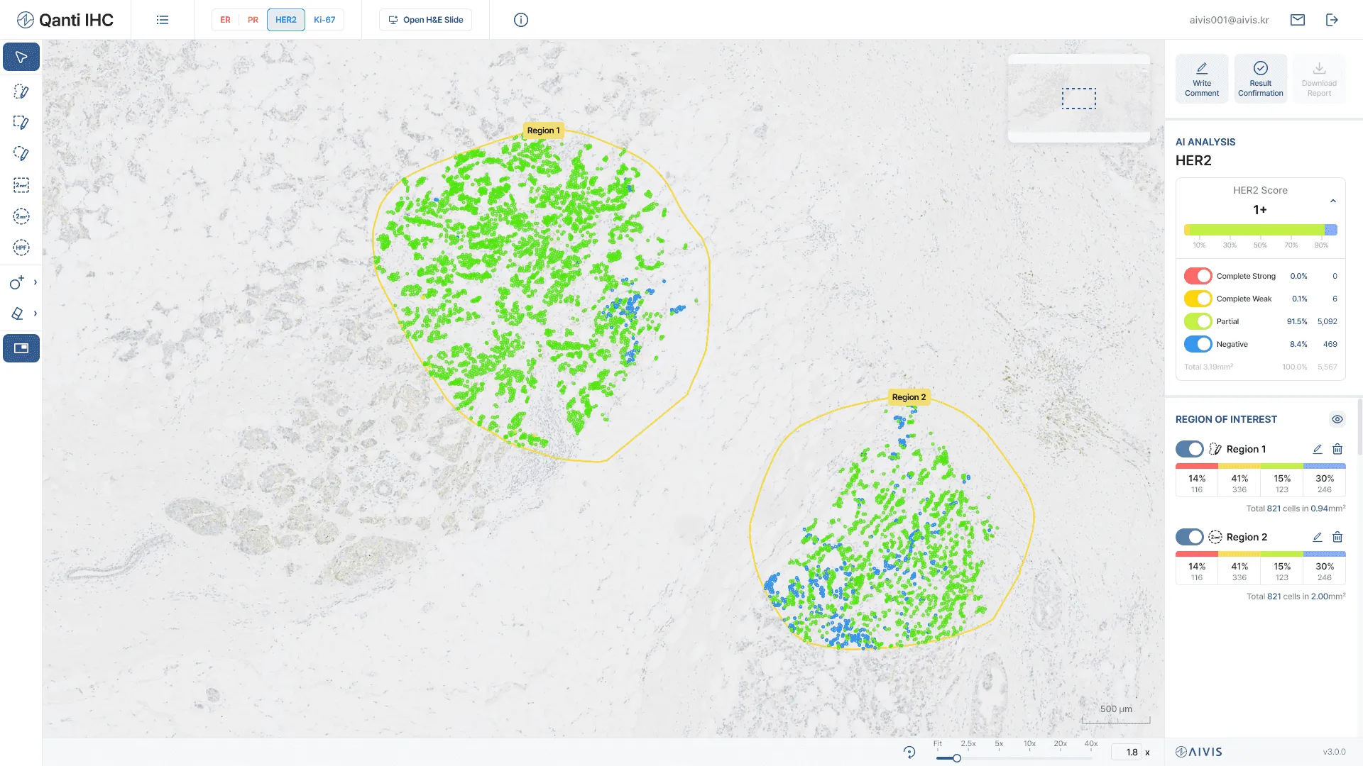
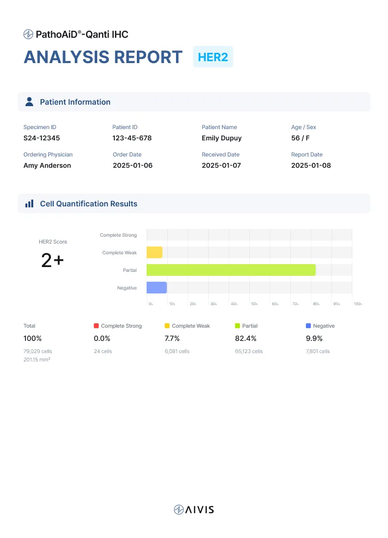
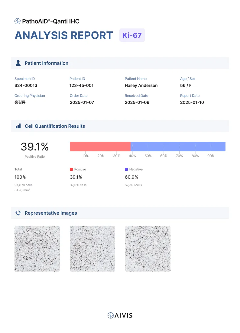
AI Based Automatic Quantification of ER, PR, HER2 and Ki-67
Fast and Reliable Performance
Qanti IHC analyzes each WSI in just 5 minutes.
Check your result within seconds after drawing region-of-interest (ROI).
Trained and validated on high-quality data from 10 in-house pathologists.
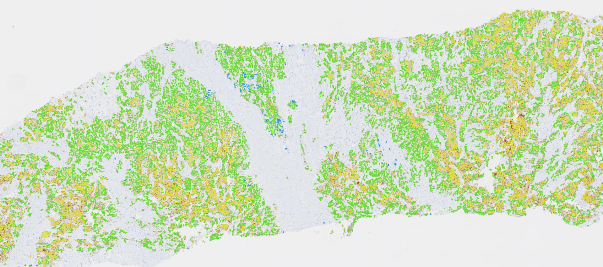
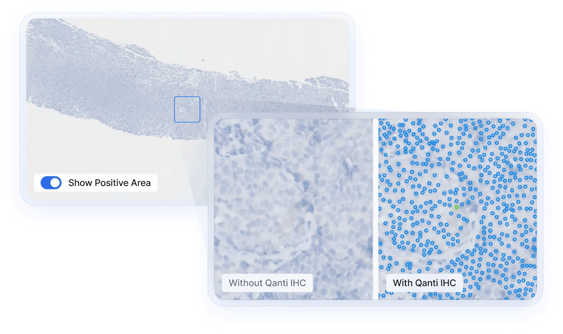
Detecting entire cells instead of only nuclei to prevent overcounting.
Pathologist-Friendly Usability and Design
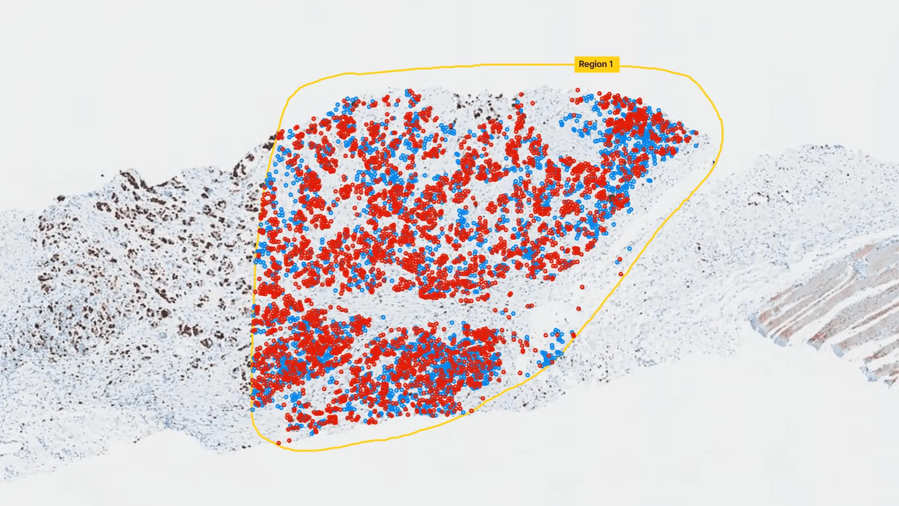
Quick and Efficient Search for Positive Cells
Qanti IHC can locate areas containing IHC-positive cells.
Save your time and effort in identifying eligible patients for target therapy.
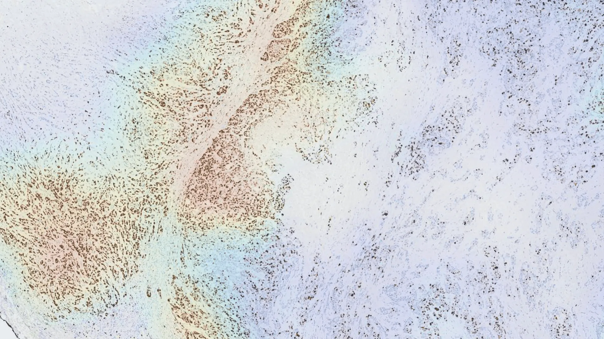
Hotspot Visualization
Qanti IHC displays IHC-positive cell distribution as a heatmap.
Quickly identify ‘hotspots’ for precise risk assessment.
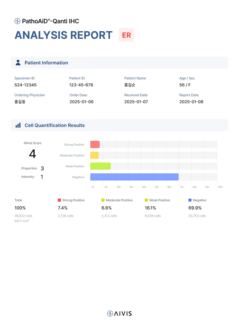
One-Click Report Generation
Qanti IHC generates a standardized quantification report based on ASCO/CAP guidelines.
Review and customize the results as needed.
Fast
Processes each whole slide image (WSI) in under 5 minutes.
Get ready-made results within seconds after drawing a region of interest (ROI).
Reliable
Trained and validated by a team of 10 full-time in-house pathologists.
Extensively tested on slides from diverse scanners and sources.
Flexible
Capable of visualizing over 3,000,000 cells at once.
Performs WSI-level cell proportion calculations for comprehensive quantification.
Pathologist-Friendly
Easy to view and modify results
Standardized ROI drawing tools, including 2mm² and HPF (high-power field) area stamps.
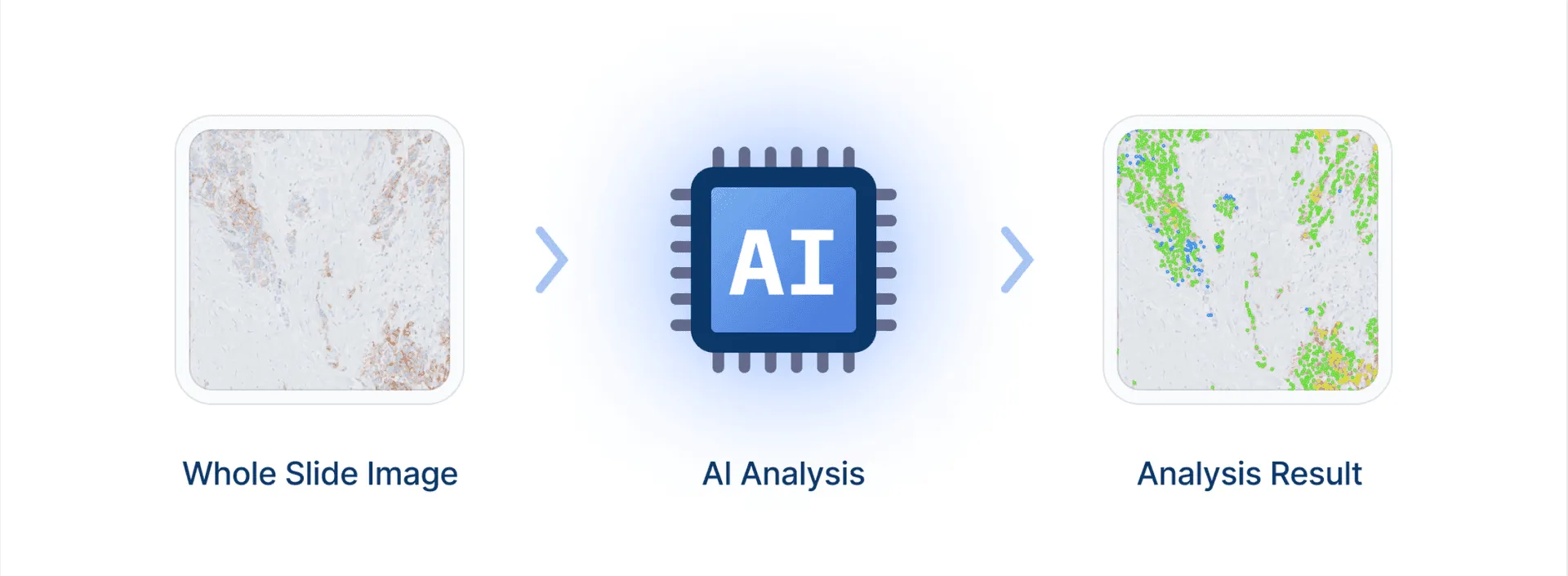
WORKFLOW
Only 5 minutes
for surgical specimen